
Recommendation
Sexual antagonism (SA), wherein the fitness interests of the sexes do not align, is inherent to organisms with two (or more) sexes. SA leads to intra-locus sexual conflict, where an allele that confers higher fitness in one sex reduces fitness in the other [1, 2]. This situation leads to what has been referred to as "gender load", resulting from the segregation of SA alleles in the population. Gender load can be reduced by the evolution of sex-specific (or sex-biased) gene expression. A specific prediction is that gene-duplication can lead to sub- or neo-functionalization, in which case the two duplicates partition the function in the different sexes. The conditions for invasion by a SA allele differ between sex-chromosomes and autosomes, leading to the prediction that (in XY or XO systems) the X should accumulate recessive male-favored alleles and dominant female-favored alleles; similar considerations apply in ZW systems ([3, but see 4].
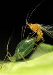
Aphids present an interesting special case, for several reasons: they have XO sex-determination, and three distinct reproductive morphs (sexual females, parthenogenetic females, and males). Previous theoretical work by the lead author predict that the X should be optimized for male function, which was borne out by whole-animal transcriptome analysis [5].
Here [6], the authors extend that work to investigate “tissue”-specific (heads, legs and gonads), sex-specific gene expression. They argue that, if intra-locus SA is the primary driver of sex-biased gene expression, it should be generally true in all tissues. They set up as an alternative the possibility that sex-biased gene expression could also be driven by dosage compensation. They cite references supporting their argument that "dosage compensation (could be) stronger in the brain", although the underlying motivation for that argument appears to be based on empirical evidence rather than theoretical predictions.
At any rate, the results are clear: all tissues investigated show masculinization of the X. Further, X-linked copies of gene duplicates were more frequently male-biased than duplicated autosomal genes or X-linked single-copy genes.
To sum up, this is a nice empirical study with clearly interpretable (and interpreted) results, the most obvious of which is the greater sex-biased expression in sexually-dimorphic tissues. Unfortunately, as the authors emphasize, there is no general theory by which SA, variable dosage-compensation, and meiotic sex chromosome inactivation can be integrated in a predictive framework. It is to be hoped that empirical studies such as this one will motivate deeper and more general theoretical investigations.
References
[1] Rice WR, Chippindale AK (2001) Intersexual ontogenetic conflict. Journal of Evolutionary Biology 14: 685-693. https://doi.org/10.1046/j.1420-9101.2001.00319.x
[2] Bonduriansky R, Chenoweth SF (2009) Intralocus sexual conflict. Trends Ecol Evol 24: 280-288. https://doi.org/10.1016/j.tree.2008.12.005
[3] Rice WR. (1984) Sex chromosomes and the evolution of sexual dimorphism. Evolution 38: 735-742. https://doi.org/10.1086/595754
[4] Fry JD (2010) The genomic location of sexually antagonistic variation: some cautionary comments. Evolution 64: 1510-1516. https://doi.org/10.1111%2Fj.1558-5646.2009.00898.x
[5] Jaquiéry J, Rispe C, Roze D, Legeai F, Le Trionnaire G, Stoeckel S, et al. (2013) Masculinization of the X Chromosome in the Pea Aphid. PLoS Genetics 9. https://doi.org/10.1371/journal.pgen.1003690
[6] Jaquiéry J, Simon J-C, Robin S, Richard G, Peccoud J, Boulain H, Legeai F, Tanguy S, Prunier-Leterme N, Le Trionnaire G (2022) Masculinization of the X-chromosome in aphid soma and gonads. bioRxiv, 2021.08.13.453080, ver. 4 peer-reviewed and recommended by Peer Community in Evolutionary Biology. https://doi.org/10.1101/2021.08.13.453080
DOI or URL of the preprint: https://doi.org/10.1101/2021.08.13.453080
Version of the preprint: 2
I only have one very specific criticism, which I don't think warrants additional review, but it does require either a rebuttal from the authors or a modest revision, which would not require additional work, only additional thinking and/or scholarship. See attached.
Download recommender's annotationsDOI or URL of the preprint: https://doi.org/10.1101/2021.08.13.453080
Dear authors,
Thank you for submitting your preprint "Masculinization of the X-chromosome in aphid soma and gonads" to PCI Evol Biol. Your manuscript has been read by two reviewers, whose comments are enclosed. As you will see, the reviews are largely positive, and, based on these reviews as well as my own evaluation, I would recommend your manuscript to be eventually included in PCI Evol Biol. However, before reaching a final decision, I would ask you to revise your manuscript according to the recommendations by the reviewers. Please address the main highlighted points, including:
- Addressing the issue of using a cut-off of 70% for deciding if a gene is morph or tissue specific (or specific to tissues within morphs). Reviewer#2 suggests using different cut-offs. An alternative would be to use a quantification of specificity for each gene (eg. across the 9 sample types) and then testing for enrichment of highly specific genes in different conditions. A good quantification for specificity is for example Tau (see Yanai et al. 2004. Genome-wide midrange transcription profiles reveal expression level relationships in human tissue) specification. Bioinformatics 21:650-659)
-Contrary to reviewer#1, I do not see the lack of single-cell resolution of gonads as a fundamental problem for this study. It might be if the X was enriched for female-biased genes (a pattern which can be mimicked by X-inactivation in male gonads), but is difficult to use as an explanation here since the X is enriched for male-biased genes and there is no evidence for reduced expression of the X in females. Nevertheless, the authors may want to extend their current discussion of how tissue allometries or different cell types within composite tissues can affect interpretations of dosage compensation in their study.
In addition to the points raised by the reviewers, I suggest investigating sex-biased expression and dosage compensation along the X chromosome. Are there any clusters of strongly male-biased genes? I also find the lack of (detected) expression of a large portion of X-linked genes overall quite interesting and suggest the authors include a discussion of this finding in their ms. Is this because X-linked genes are generally lowly expressed (so they do not pass the filtering) or highly tissue-specific (for tissues not present in the sequenced body fragments)? Are they detected in the whole-body samples in the previous studies? Furthermore, generating strongly male-biased expression of X-linked genes in an XX/X0 species will require extreme up-regulation in males. Is there evidence for increased gene duplication on the X relative to autosomes (with both/all copies located on the X)? Such a pattern could facilitate strong expression from a single X.
I also encourage the authors to revise their manuscript according to the more minor suggestions from the reviewers, which will certainly improve it. To additional minor comments:
I suggest reformulating “By contrast, segregation distortion cannot affect the aphid X chromosome during spermatogenesis, as all sperm cells that do not carry an X degenerate.”. This can be considered the most extreme form of segregation distortion (100% transmission instead of the expected 50%).
Please tone down “Here, we predicted that if masculinization of the aphid X chromosome evolved solely in response to intra-locus sexual conflicts, masculinization would occur in all tissues.” to “a strong driver of” or something like this (“soley” would require excluding other factors, which I do not think is the authors intention. Furthermore there is no direct evidence that genes with male-biased expression are actually generally beneficial for males, even though this is certainly an implicit assumption in many studies)
Best regards,
Tanja Schwander
In this study, the authors predicted that if masculinization of the pea aphid X chromosome evolved solely in response to intra-locus sexual conflicts, masculinization would occur in all tissues. To verify this prediction they used chromosome-scale assembly and bulk RNA-seq of different sexes/tissues/morphs of the pea aphid to measure gene expression levels of the X and autosomes, across samples. They found masculinization of the X in each type of tissue. Furthermore, authors stated that the X-linked copy of a duplicated gene is more likely to show a male-biased expression than its autosomal copy or an X-linked single copy gene. The findings suggest that duplications facilitate sub or neofunctionalization toward the sex-specific optimum. Overall, the ms is an extension of prevoius theoretical and empirical works from some of the authors supporting the hypothesis of that the large excess of male-biased genes observed on the pea aphid X chromosome compared to autosomes has evolved in response to sexual conflicts, by restricting the product of a sexually antagonistic allele to the sex it benefits. These observations provide new information about the atypical genome-wide pattern of gene expression in pea aphid, with a high degree of masculinization of the X chromosome in both somatic and gonadic tissues. The methodology and the details of the experiments are clearly described and appropriate. I have only some minor concerns that I think the authors should address before the ms progress it further.
#1. A major potential caveat of this study is the expression level of the X chromosome during spermatogenesis. Germ cells enact a very specialized program of differentiation and bulk RNA-seq alone is not enough to accurately determine the transcriptional profile of the X chromosome along the spermatogenesis. Two recent Drosophila papers that used single-cell RNA-seq experiments (see below) have showed that the X undergone dosage compensation earlier in spermatogenesis and inactivation in secondary spermatocytes. Folowing this idea, the masculinization of the X chromosomes (or the male-biased gene expression) in pead aphid may be because dosage compensation in spermatogonia; however the picture could be different during the transition to meiosis. That being said, how authors exclude this posiblity uisng bulk RNA expression alone? There may be other ways to do this too, but the current approach does not seem sufficient. Furthermnore, previous indiret evidences in other X0 systems (e.g. crickets and grasshoppers) have suggested that the X chromosome is inactived during male meioses similar to the XY body in mammals because of the differential chromathin condensation patthern of the X (called heteropicnosis) regarding autosomes.
#2 Page 18 (Discussion), middle of the first paragraph:
- It said that "Indeed, sex chromosomes of other dosage-compensated species are generally not compensated in the gonads of the heterogametic sex in diptera and lepidoptera (Vicoso and Bachtrog 2015; Gu et al. 2017; Gu and Walters 2017)". I would suggest not generalizing the idea. Using single-cell RNA-sequencing, two recent Drosophila paper (https://doi.org/10.1038/s41467-021-20897-y; https://journals.plos.org/plosgenetics/article?id=10.1371/journal.pgen.1009728) have showed that X chromosome is expressed at a higher rate in spermatogonia than expected based on DNA copy number alone, supporting the idea of X chromosome dosage compensation in the premeiotic male germline. However, authors also showed that the X chromosome is specifically inactivated in primary spermatocytes (MSCI) based on measuring all genes or a set of broadly expressed genes in testis they identified (https://doi.org/10.1038/s41467-021-20897-y). Please correct this sentence accordengly.
#3 Page 18 (Discussion), at the end of the first paragraph:
- It is said that "The silencing of sex-linked genes during gametogenesis could have evolved to protect from the invasion of segregation distorters that would bias sex-ratio (Meiklejohn and Tao 2010)". Other transcriptional silencing mechanisms can evolve to protect the largely dissimilar XY or X0 chromosomes by defending un-synapsed regions from stealthy invasive transposons that cannot be detected as foreign in the absence of a homolog. Silencing may also protect against unwanted recombination between the X and Y, or to repair damage created by lack of recombination in XY or X0. I would suggest that the authors add these potential mechanisms in the text, and stress that these models are unlikely in the case of aphid males X0, which completely lack meiotic recombination.
In this paper, the authors test in the pea aphid whether masculinisation of the X chromosome is present in all tissues or restricted to only a few. In the latter case, sexual antagonism cannot explain the distribution of sex-biased genes as this presumably affects all tissues equally. They find that the enrichment of male-biased genes concerns all tissues and the authors take this as further support that sexual antagonism together with the interesting mode of reproduction (XX/X0 and cyclic parthenogenesis) drive the masculinisation of the X in this system.
The study is nicely designed and carried out. All the ground work on sex-biased genes in pea aphid has already been laid by previous work by Jaquiéry et al. and it is a natural next step to look at individual tissues (or compound structure as it is mostly the case here) to if this is maybe driven by individual tissues such as the gonads or the brain as has been found in other systems.
I also like that the authors also look at duplicated genes that can alleviate sexual antagonism and in a system with sex chromosomes, can also be translocated to either the autosomes or the sex chromosomes (in this case only the X). This study represents an interesting case where it is indeed possible to show that specifically male-biased copies are retained on or translocated to the X.
The one major point in the methodology that I find strange is the definition of biased genes. Why do the authors use 70% of all reads as a cut-off? At least it should be demonstrated that 60% or 80% does deliver the same patterns. This cut-off seems somewhat arbitrary to me and is not explained. Furthermore, what happens if you instead use traditional differential expression analysis? Do the patterns hold and you just loose statistical power because of fewer genes in your analysis or does this change any thing more fundamentally? Because the programmes do have a point of throwing out very lowly expressed genes or those with strong heterogeneity among replicates. As the whole paper builds on this analysis, I think it is crucial to clarify this point, i.e. explain why use 70% and show that it does not change the results qualitatively if another method to define differential expression is used.
In addition, I have only a few minor comments that could be rather easily addressed:
1) Abstract: “We observed a masculinization of the X at the tissue-level, with male-biased genes being 2.5 to 3.5 more frequent on the X than expected.”
This is not quite clear. Upon first reading, I thought this meant that masculinisation is only observed in some tissues.
2) I think there is a typo/mistake here in the parenthesis, at least it is not clear to me what the “whether sexual or parthenogenetic” means:
“The mode of Log2 ratio of male-to-female log2(RPKM+1) (whether sexual or parthenogenetic, figure 4CFI and Supplementary figure S2) lies close to 0 for both autosomal and X-linked genes”
3) “This indicates an overexpression of some of the genes located on the single X chromosome of male cells, which exceeds dosage compensation. This pattern was expected, given that male-biased genes are significantly more frequent on the X than on autosomes (figure 2).”
So when sex-biased genes (more than 2-fold different) are excluded, is there full dosage compensation? This point was not entirely clear to me and should be tested as the enrichment of highly male-biased genes could mask the underlying patterns.
4) While in the figures, I find the abbreviation for the sample types helpful, they reduce readability in the text a bit. Maybe it makes sense to write “parthenogenetic female legs” rather than “PL” for example here: “Conversely, parthenogenetic female-biased genes were significantly less frequent on the X, except for PL+ genes due to lack of power[...]”
5) Concerning dosage compensation of the testes in the discussion: Is it known whether the X chromosome has fewer TEs than other arthropods where X-inaktivation or X-downregulation occurs?
6) I do not understand this part of the methods: “Given the low number of genes satisfying this criterion (n=210), gene classes with male-biased expression (i.e., M+, MG+, ML+ and MH+ genes) were aggregated. ” What do you mean by aggregated? Did you use additional genes, i.e. ones other than those with 1 copy on the X and 1 copy on an autosome?
