Effect of sex chromosomes on mammalian behaviour: a case study in pygmy mice
 based on reviews by Marion Anne-Lise Picard, Caroline Hu and 1 anonymous reviewer

Genotypic sex shapes maternal care in the African Pygmy mouse, Mus minutoides
Abstract
Recommendation: posted 26 September 2022, validated 31 October 2022
Marais, G. and Bilde, T. (2022) Effect of sex chromosomes on mammalian behaviour: a case study in pygmy mice. Peer Community in Evolutionary Biology, 100152. https://doi.org/10.24072/pci.evolbiol.100152
Recommendation
In mammals, it is well documented that sexual dimorphism and in particular sex differences in behaviour are fine-tuned by gonadal hormonal profiles. For example, in lemurs, where female social dominance is common, the level of testosterone in females is unusually high compared to that of other primate females (Petty and Drea 2015).
Recent studies however suggest that gonadal hormones might not be the only biological factor involved in establishing sexual dimorphism, sex chromosomes might also play a role. The four core genotype (FCG) model and other similar systems allowing to decouple phenotypic and genotypic sex in mice have provided very convincing evidence of such a role (Gatewood et al. 2006; Arnold and Chen 2009; Arnold 2020a, 2020b). This however is a new field of research and the role of sex chromosomes in establishing sexually dimorphic behaviours has not been studied very much yet. Moreover, the FCG model has some limits. Sry, the male determinant gene on the mammalian Y chromosome might be involved in some sex differences in neuroanatomy, but Sry is always associated with maleness in the FCG model, and this potential role of Sry cannot be studied using this system.
Heitzmann et al. have used a natural system to approach these questions. They worked on the African Pygmy mouse, Mus minutoides, in which a modified X chromosome called X* can feminize X*Y individuals, which offers a great opportunity for elegant experiments on the effects of sex chromosomes versus hormones on behaviour. They focused on maternal care and compared pup retrieval, nest quality, and mother-pup interactions in XX, X*X and X*Y females. They found that X*Y females are significantly better at retrieving pups than other females. They are also much more aggressive towards the fathers than other females, preventing paternal care. They build nests of poorer quality but have similar interactions with pups compared to other females. Importantly, no significant differences were found between XX and X*X females for these traits, which points to an effect of the Y chromosome in explaining the differences between X*Y and other females (XX, X*X). Also, another work from the same group showed similar gonadal hormone levels in all the females (Veyrunes et al. 2022).
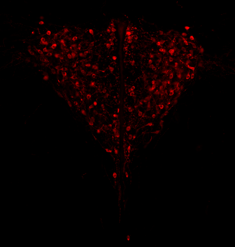
Heitzmann et al. made a number of predictions based on what is known about the neuroanatomy of rodents which might explain such behaviours. Using cytology, they looked for differences in neuron numbers in the hypothalamus involved in the oxytocin, vasopressin and dopaminergic pathways in XX, X*X and X*Y females, but could not find any significant effects. However, this part of their work relied on very small sample sizes and they used virgin females instead of mothers for ethical reasons, which greatly limited the analysis.
Interestingly, X*Y females have a higher reproductive performance than XX and X*X ones, which compensate for the cost of producing unviable YY embryos and certainly contribute to maintaining a high frequency of X* in many African pygmy mice populations (Saunders et al. 2014, 2022). X*Y females are probably solitary mothers contrary to other females, and Heitzmann et al. have uncovered a divergent female strategy in this species. Their work points out the role of sex chromosomes in establishing sex differences in behaviours.
References
Arnold AP (2020a) Sexual differentiation of brain and other tissues: Five questions for the next 50 years. Hormones and Behavior, 120, 104691. https://doi.org/10.1016/j.yhbeh.2020.104691
Arnold AP (2020b) Four Core Genotypes and XY* mouse models: Update on impact on SABV research. Neuroscience & Biobehavioral Reviews, 119, 1–8. https://doi.org/10.1016/j.neubiorev.2020.09.021
Arnold AP, Chen X (2009) What does the “four core genotypes” mouse model tell us about sex differences in the brain and other tissues? Frontiers in Neuroendocrinology, 30, 1–9. https://doi.org/10.1016/j.yfrne.2008.11.001
Gatewood JD, Wills A, Shetty S, Xu J, Arnold AP, Burgoyne PS, Rissman EF (2006) Sex Chromosome Complement and Gonadal Sex Influence Aggressive and Parental Behaviors in Mice. Journal of Neuroscience, 26, 2335–2342. https://doi.org/10.1523/JNEUROSCI.3743-05.2006
Heitzmann LD, Challe M, Perez J, Castell L, Galibert E, Martin A, Valjent E, Veyrunes F (2022) Genotypic sex shapes maternal care in the African Pygmy mouse, Mus minutoides. bioRxiv, 2022.04.05.487174, ver. 4 peer-reviewed and recommended by Peer Community in Evolutionary Biology. https://doi.org/10.1101/2022.04.05.487174
Petty JMA, Drea CM (2015) Female rule in lemurs is ancestral and hormonally mediated. Scientific Reports, 5, 9631. https://doi.org/10.1038/srep09631
Saunders PA, Perez J, Rahmoun M, Ronce O, Crochet P-A, Veyrunes F (2014) Xy Females Do Better Than the Xx in the African Pygmy Mouse, Mus Minutoides. Evolution, 68, 2119–2127. https://doi.org/10.1111/evo.12387
Saunders PA, Perez J, Ronce O, Veyrunes F (2022) Multiple sex chromosome drivers in a mammal with three sex chromosomes. Current Biology, 32, 2001-2010.e3. https://doi.org/10.1016/j.cub.2022.03.029
Veyrunes F, Perez J, Heitzmann L, Saunders PA, Givalois L (2022) Separating the effects of sex hormones and sex chromosomes on behavior in the African pygmy mouse Mus minutoides, a species with XY female sex reversal. bioRxiv, 2022.07.11.499546. https://doi.org/10.1101/2022.07.11.499546
The recommender in charge of the evaluation of the article and the reviewers declared that they have no conflict of interest (as defined in the code of conduct of PCI) with the authors or with the content of the article. The authors declared that they comply with the PCI rule of having no financial conflicts of interest in relation to the content of the article.
This work was supported by the French National Research Agency (ANR grant SEXREV 18-CE02-0018-01)
Evaluation round #2
DOI or URL of the preprint: https://doi.org/10.1101/2022.04.05.487174
Version of the preprint: 2
Author's Reply, 19 Sep 2022
We posted the final version on the preprint server
Decision by Gabriel Marais and Trine Bilde , posted 09 Sep 2022
The referees were positive about the revised ms but all asked for some minor changes. Could you please revise your ms accordingly, and we will be happy to recommend your preprint.
Reviewed by anonymous reviewer 1, 17 Aug 2022
Reviewed by Caroline Hu, 31 Aug 2022
Overall I think this preprint has reached a quality level suitable for recommendation. The authors have made significant changes to the manuscript that improved it in the following ways:
1. Clearer motivations and hypotheses for cell population comparison
2. Appropriately conservative interpretation of histology results
3. Increased detail in methods
4. Much more legible figures
Figure suggestions:
Figure 1 – The two boxed texts “1 - Candidate neural…” and “2 - Predictions…” would serve better as “headers” for the upper and lower half of the figure if they were placed more towards the top-left or top-center. If you “read” the figure like you would text (left to right, top to bottom), these headers are a bit buried.
Figure 3 – I suggest moving the Mus atlas image and the “c” label to the left of the PVN images.
Reviewed by Marion Anne-Lise Picard, 05 Sep 2022
This new version of the manuscript from L. Heitzmann et al. fulfill my previous suggestions :
Particularly, they have added a new Figure 1 that very well describes the investigated neural circuits, and clarifies their predictions. It greatly improves the comprehension!
Importantly, the authors now state that all female genotypes display similar gonadal/adrenal hormonal levels (lines 387 to 390) - and they added the corresponding reference (Veyrunes et al. 2022).
The authors did not retain the idea of comparing left side vs right side of the hypothalamus, but this suggestion was not mandatory, as it does not affect the conclusions of their paper.
The authors improved the readability of Figure 2 and completed the caption of Fig. S6.
All typos were corrected.
Evaluation round #1
DOI or URL of the preprint: https://doi.org/10.1101/2022.04.05.487174
Version of the preprint: 1
Author's Reply, 15 Jul 2022
Decision by Gabriel Marais and Trine Bilde , posted 27 Jun 2022, validated 27 Jun 2022
Dear authors,
Three referees have seen your preprint and made a number of comments/suggestions, which we think will help you improve it.
Could you please submit a revised version and a rebuttal adressing all referees' comments (see PCI Evol Biol guidelines)?
In particular:
- I agree with referee 3 that clear predictions about the different experiments should be provided in the introduction
- Following one comment of referee 3 (about discussion), I think showing a working model (figure?) of the effects of sex chromosomes on the studied phenotypes (with potential specific roles of XCI escapees - see referees 2 and 3, etc...) would be useful
- I found referee 1 suggestion on analysing separately data from right and left parts of hypothalamus interesting, please consider it.
Best regards,
Reviewed by Marion Anne-Lise Picard, 26 May 2022
# SUMMARY OF THE MANUSCRIPT:
In this study, L. Heitzmann et al. investigate the role of the sex chromosome complement on maternal care, by combining behavioural and neurobiological aspects. The studied model, Mus minutoides, is characterized by three distinct female genotypes: XX, XX* and X*Y, all occurring naturally. Males are XY. This allow them not only to disentangle the role of gonadal hormone (phenotypic sex) vs sex chromosomes (genotypic sex); but also to discuss their results in the light of natural selection.
First, they performed an exhaustive behavioural assay, with a high number of replicates (from 75 to 105 females), and described as main results:
(i) higher pup retrieval efficiency in X*Y females,
(ii) no change in mother-pup interactions,
(iv) poor nest quality in X*Y females,
(v) no biparental care in X*Y females that are highly aggressive towards males.
Then, they focus on neural circuit known to underly such behaviors in model species, and report:
(i) no significant differences in oxytocin expressing neurons number
(ii) no significant differences in vasopressin expressing neurons number, but a tendancy for lower number in X*Y females
(iii) no significant differences in the density of tyrosine hydroxylase expressing neurons, but a tendancy for greater density in X*Y females
They conclude that the sex chromosome complement shapes sexually dimorphic behaviours, and that different combinations of sex chromosomes lead to alternative parental care strategies (more than the two expected under the sole effect of gonadal hormones). By providing the first description of the brain structure for the four genotypes of this species, they also open new perspectives for the study of the neural bases of such behaviours.
# MY OVERALL IMPRESSION:
That manuscript is straightforward and well written. It is attracting for a wide audience interested in reproductive biology, ranging from parental care, to the role of sex chromosomes in shaping phenotypic differences between sexes. I recommend it for three main reasons: 1. First, they shuffle the long standing thought that gonadal hormones are the main driver of sexually dimorphic behaviors; 2. their model, characterized by naturally occuring sex reversal, is original and relevant; 3. they propose complementary approaches.
# MINOR SUGGESTIONS:
1. The only important concern I could have is that their model do not allow them to perform the neurobiology assays on mothers, but on virgin females instead. Nonetheless, they adequately discuss this limitation (l390-l406) and their arguments are valuables. Even if I understand the reasoning behind the formulation "the neural basis of maternal behaviours", they maybe could find an alternative which better reflects the "potentiality"?
2. Sup.Fig. S5-S10: Why not comparing the "oxytocin neurons distribution" (S6) in the same way as vasopressin neurons (S5)? (It seems that at least that the regions #1 and #5 are also characterised by a tendancy for lower number of oxytocin neurons, aren't they?)
Besides, did the autor studied the distribution of cell on left vs right side??
Indeed, there are growing evidences that the hypothalamus is functionally lateralized (Kiss et al. Brain Science, 2020 for review : https://www.ncbi.nlm.nih.gov/pmc/articles/PMC7349050/). Such analysis may allow them to refine the signal?
3. Finally, did the authors previously assessed the level of gonadal hormones in the different female genotypes? Are they similar?
# VERY MINOR SUGGESTIONS:
-Fig2 : make slightly bigger panels A & B ?
-Even if it is well explained in the text, maybe the authors could had a small chart of the studied neural circuit?
# SOME TYPOS:
l26 (keywords) : natural sex reversal and not "sex reversal, natural," ?
l106 : remove the space before "Behavioural"
l214 : remove space before "Higher"
l352 : change the ref. (Veryunes & Perez, 2018) for (Veyrunes & Perez, 2018)
l622 & 654: remove the space after "2001)"


